-
Products
- Gas analysis systems
- GAOS SENSON gas analyzers
- GAOS MS process mass spectrometry
- MaOS HiSpec ion mobility spectrometer
- MaOS AxiSpec ion mobility spectrometer
- Applications
- News
- Events
- About us
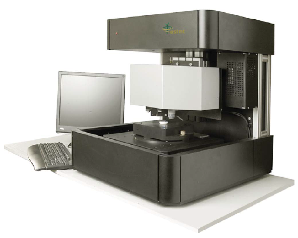
XROS MF30 – laboratory x-ray microscope-microprobe for studies of the objects by the methods of the optical microscopy, radiography, local element XRF microanalysis with the possibility of the element mapping.
Using a microscope, a sample of up to 400 mm in size along the Y-axis and of unlimited size along the X-axis (max. scan area 150×150 mm; in the case of a larger area, the scanned areas can be stitched) and up to 105 mm high can be performed.
An overview video camera and two optical microscopes with magnification up to 200 times are using for accurate determination of the scanning area.
The central optical microscope with automated sharpness adjustment is combined with the axis of the microprobe (axis of the x-ray beam).
Local X-ray fluorescence microanalysis with the possibility of elemental mapping and X-ray studies can be carried out both separately and simultaneously.
The sample positioning accuracy is 10 microns.
The minimum diameter of the x-ray probe is 30 µm.
The range of simultaneously measured elements from 11Na to 92U.
Two ancient icons were provided for the study. The image is made on a wooden canvas with a thickness of about 20 mm.
Small holes on the first icon appeared as a result of attaching the icon frame to it. The frame was removed for analysis.
The second icon shows defects in the paint layers.
![]()
|
|
|
|
|
|
|
|
|
|
|
|
|
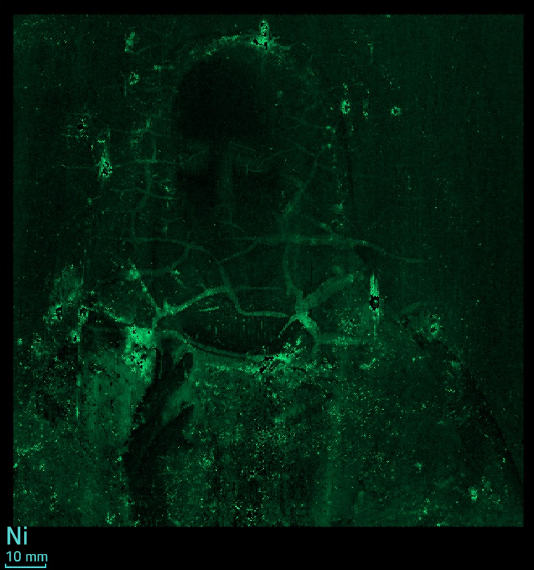
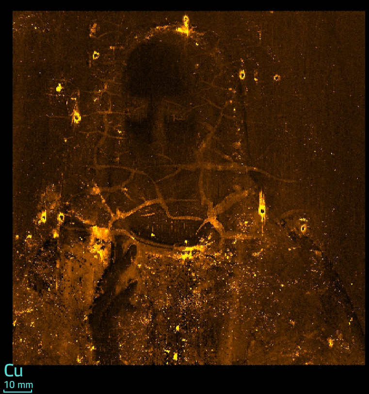
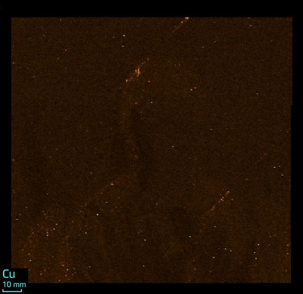
From the distribution of copper it is clear that the frame was nailed to the icon with copper nails – there is an increased copper content near the holes. The distribution shows how copper over time diffused into the wood on which the icon was painted.
![]()
|
|
|
|
|
|
|
|
|
|
|
|
|
|
|
|
|
|
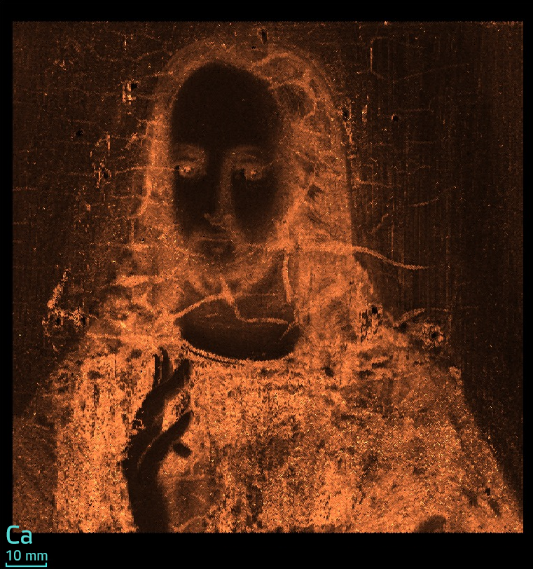
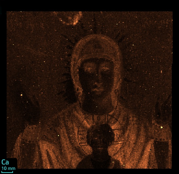
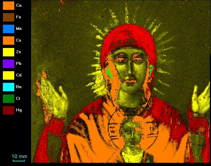
On the distribution of calcium visible outlines of the figure of the Mother of God. It can be assumed that calcium is a part of paints.
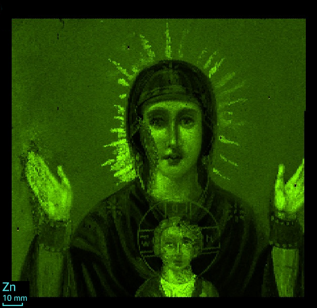
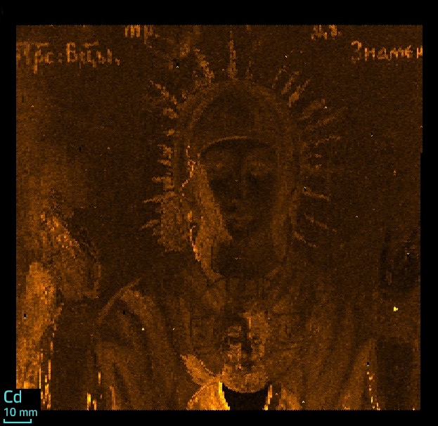
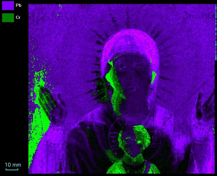
The photographs of the icon show defects in the layers of paint on the hand and face of the Mother of God. It is assumed that in these areas the paint was lost and it was restored. Probably, the boy's face was also restored, although the defects on the icon are not visualized.
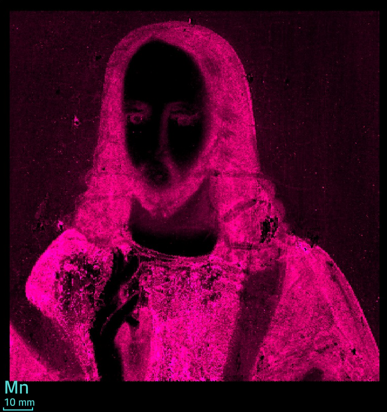
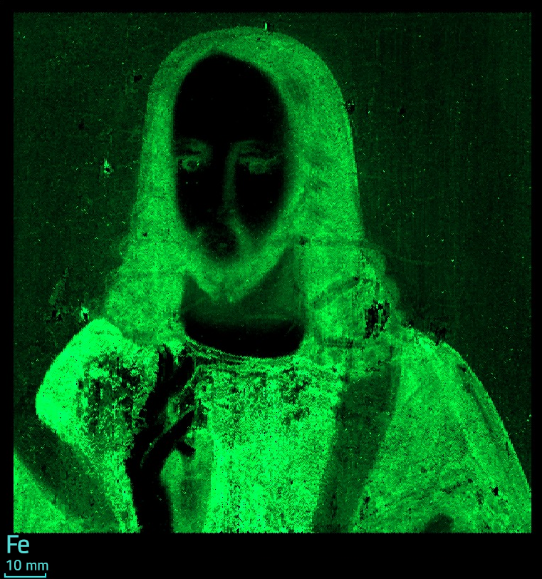
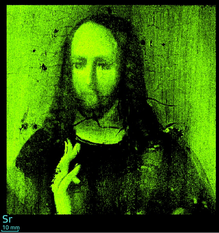
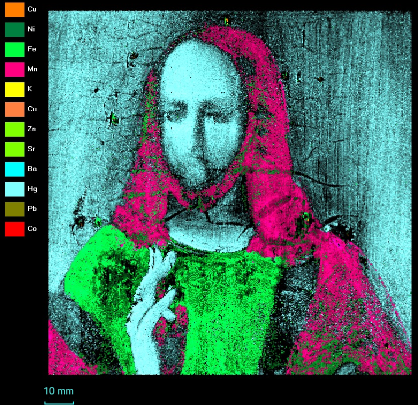
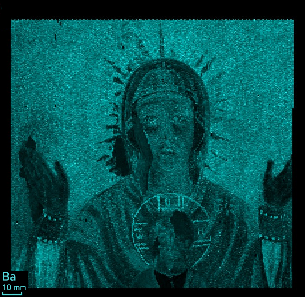
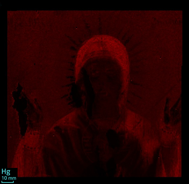
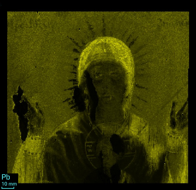
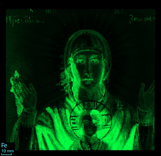
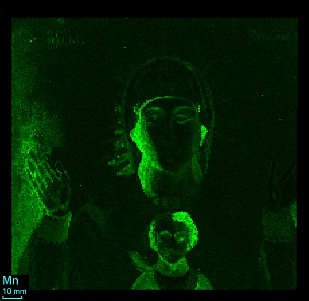
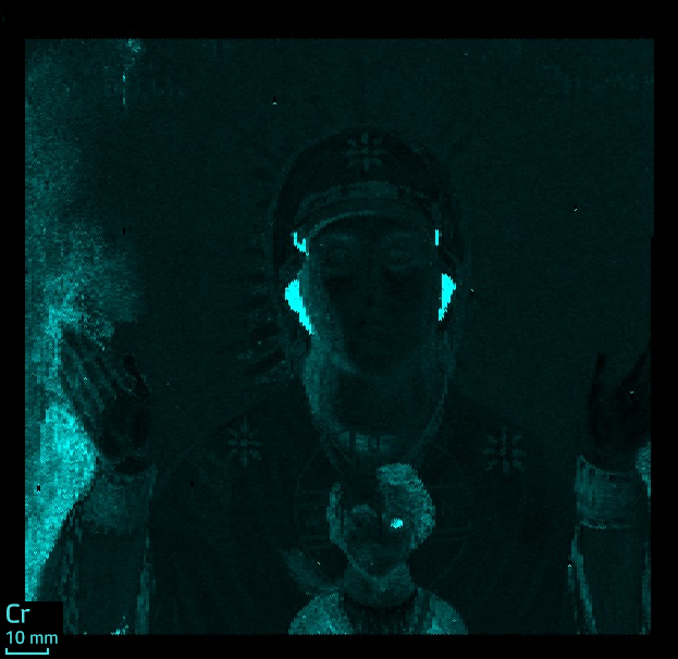
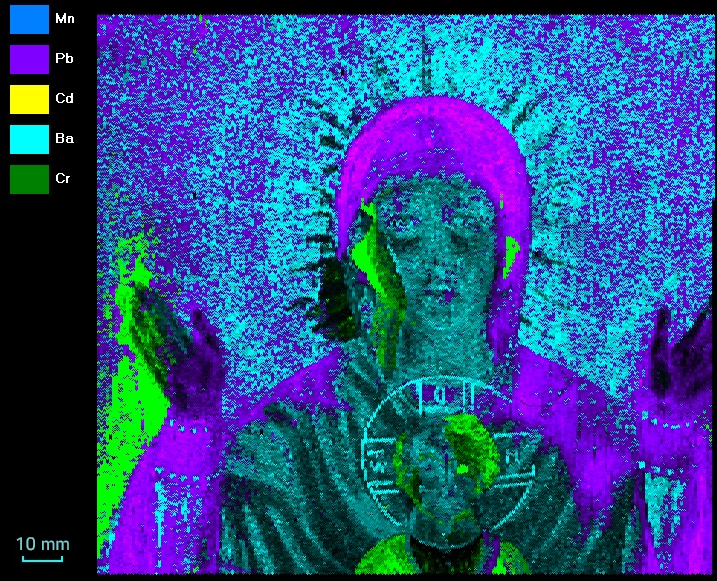
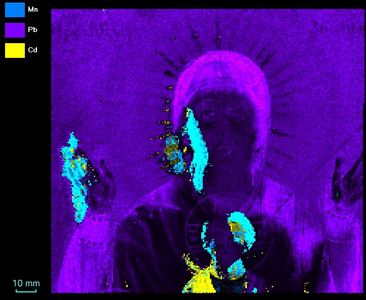
Comparing the data on all elements, it is possible to determine the sequence of applying layers of paint, as well as the course of further restoration work.
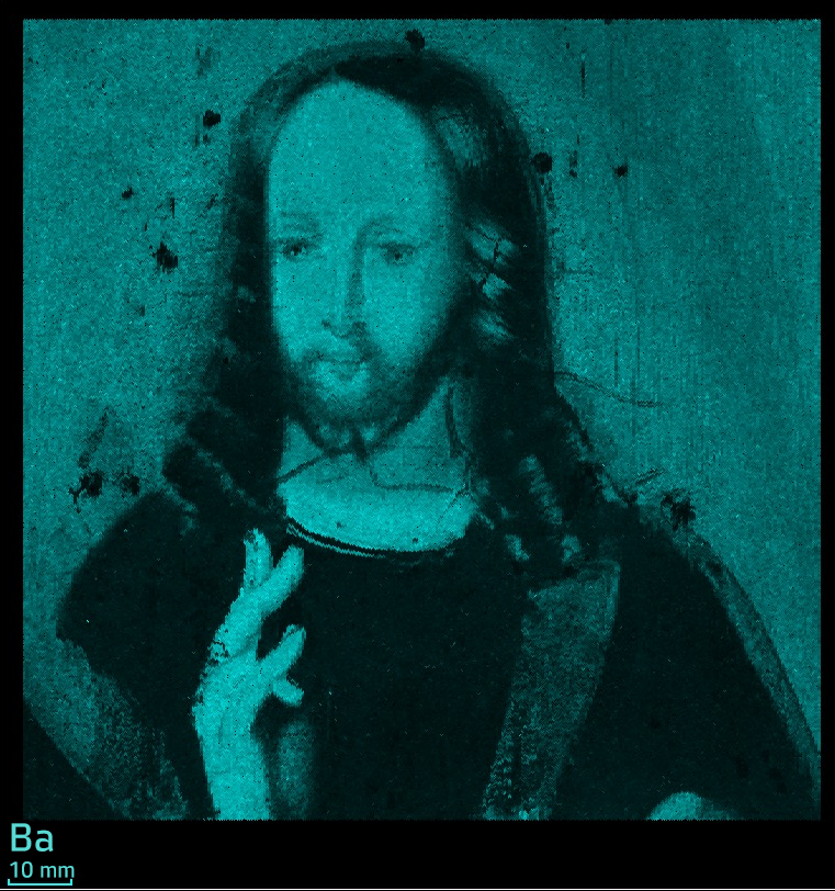
At first there was a precoating containing barium.
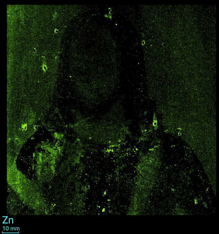
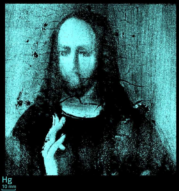
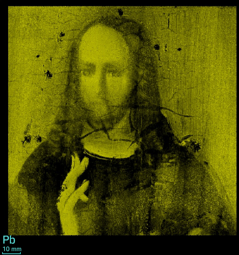
Then the figure of the Mother of God was drawт (contains in colors mercury, plumbum). Then, in places where the paint was peeling off, the background was restored with paints containing iron, chromium, and manganese. They re-painted the figure: the dress of Our Lady with paints with iron, the sleeves with barium and zinc. Left hand-painted with zinc paint. It should be noted that despite the absence of visible defects on the right hand, it was also redrawn. Cadmium and chrome are present in the colors of Jesus’s garment, and zinc in his shirt.
Then the boy's nimbus is painted with barium, zinc and iron paints, and the nimbus of our Lady is painted with zinc paints. The inscription above is painted with cadmium.
The dotted presence of copper may indicate the use of a copper frame: copper particles diffuse into paint and wood over time.
The study revealed the presence in icon paints of such elements as Ca, K, Zn, Cu, Ni, Co, Mn, Fe, Sr, Ba, Hg, Pb.
Information about the presence of certain elements in the pigments of paint helps in determining the age of the art object. And their spatial distribution gives information about the technique of writing the image - for example, the technique of multi-layer painting, technique a la prima, etc.
XROS MF30 x-ray analytical microscope-microprobe allows analyzing various art objects for the distribution of chemical elements in it with a high spatial resolution and elemental sensitivity.
|
Experiment #1 Scan step 400 µm Scan rate 400 µm/s Measurement time 1 000 ms Voltage 45 kV Electric current 5 000 µA XRT Mo anode Atmosphere Air |
Experiment #2 Scan step 700 µm Scan rate 700 µm/s Measurement time 500 ms Voltage 30 kV Electric current 6 000 µA XRT Mo anode Atmosphere Air |