-
Products
- Gas analysis systems
- GAOS SENSON gas analyzers
- GAOS MS process mass spectrometry
- MaOS HiSpec ion mobility spectrometer
- MaOS AxiSpec ion mobility spectrometer
- Applications
- News
- Events
- About us
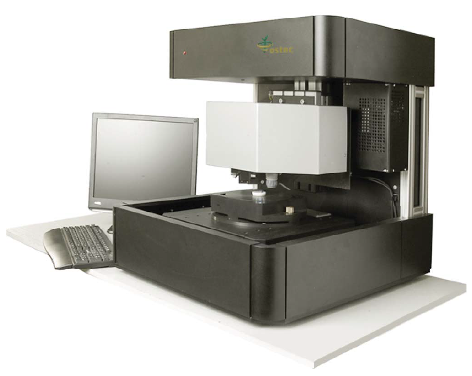
XROS MF30 – laboratory x-ray microscope-microprobe for studies of the objects by the methods of the optical microscopy, radiography, local element XRF microanalysis with possibility of the element mapping.
Using a microscope, a sample of up to 400 mm in size along the Y axis and of unlimited size along the X axis (max. scan area 150×150 mm; in the case of a larger area, the scanned areas can be stitched) and up to 105 mm high can be performed.
An overview video camera and two optical microscopes with magnification up to 200 times are using for accurate determination of the scanning area.
The central optical microscope with automated sharpness adjustment is combined with the axis of the microprobe (axis of the x-ray beam).
Local X-ray fluorescence microanalysis with the possibility of elemental mapping and X-ray studies can be carried out both separately and simultaneously.
Sample positioning accuracy is 10 microns.
The minimum diameter of the x-ray probe is 30 µm.
The range of simultaneously measured elements from 11Na to 92U.
A study of the belt onlay of rich horse harness of medieval nomads of the 10-13 century AD from archaeological excavations was carried out. The sample for research was provided by N. Kurganov, a member of the Laboratory of Conservation and Restoration of RAS IHMC.
The object of research was found during archaeological excavations of the Baidag 10 burial ground, researched by the IIMK RAS Tuva archaeological expedition. Stripes of the leather belt have completely decomposed below ground, and the onlays are very poorly preserved and covered with a thick layer of corrosion. During the restoration process, an ornament made of gold-covered silver foil was found on the surface of the iron onlays. A method used to attach the foil is remarkable: originally covered with a rhombic pattern, it was ground into the base, and then punched through with a sharp tool. As a result, the foil was covered with an additional ornament, and, thanks to the burrs forming along the edges of the holes, it was firmly held on an iron base. During several centuries below ground, the ornament that used to be magnificent became hidden under a layer of products of iron corrosion, particles of soil and mineralized organic matter. Iron used as the basic material for buckles, plates completely mineralized under the silver. The technology of production of buckles and the selection of restoration techniques is considered in detail in the article [1].
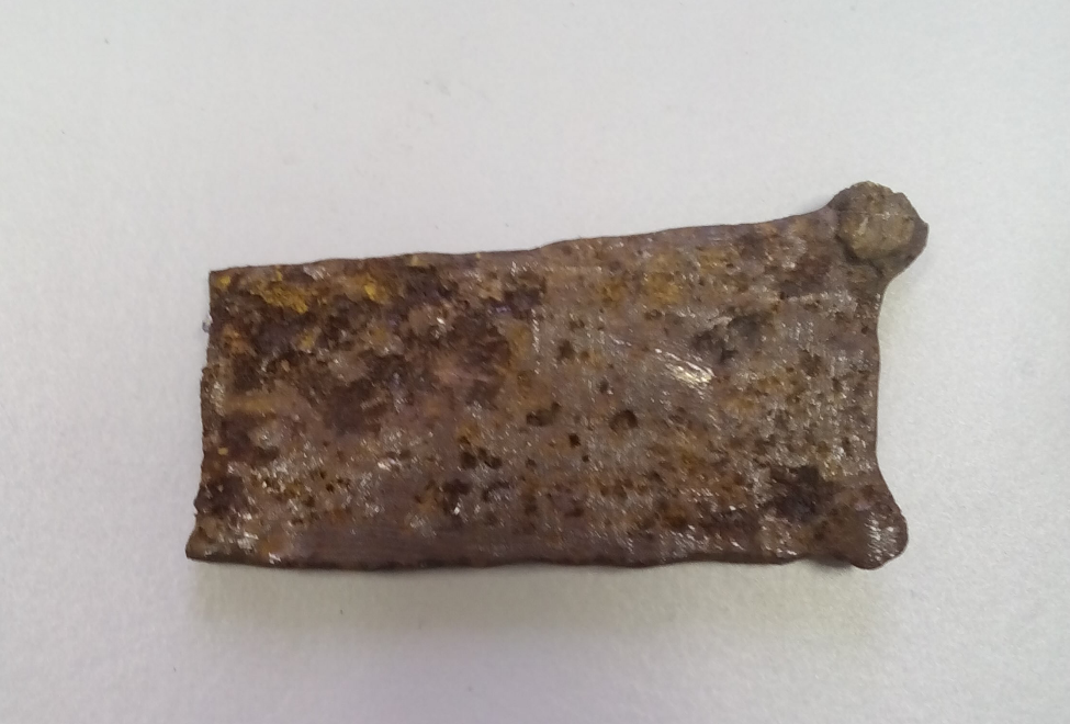
Fig. 1. Photo of a link of a nomad belt
Fig. 1 shows the appearance of the link. Fig. 2 shows the selected scan area. Fig. 3-12 show elemental mapping of the selected area of the sample. Fig. 13 shows the picture of an onlay fragment before and after restoration.
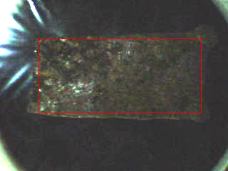
Fig. 2. Belt link. Red highlights the scan area
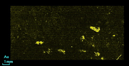
Fig. 3. Distribution of gold
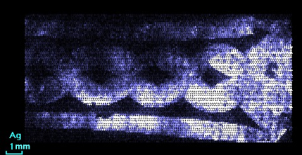
Fig. 4. Distribution of silver
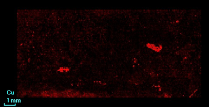
Fig. 5. Distribution of copper
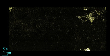
Fig. 6. Distribution of calcium
![]()
Fig. 7. Distribution of silicon
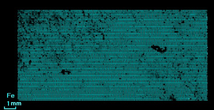
Fig. 8. Distribution of iron
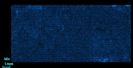
Fig. 9. Distribution of manganese
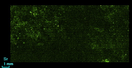
Fig. 10. Distribution of strontium
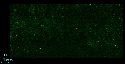
Fig. 11. Distribution of titanium

Fig. 12. Distribution of silver (green) and gold (yellow)
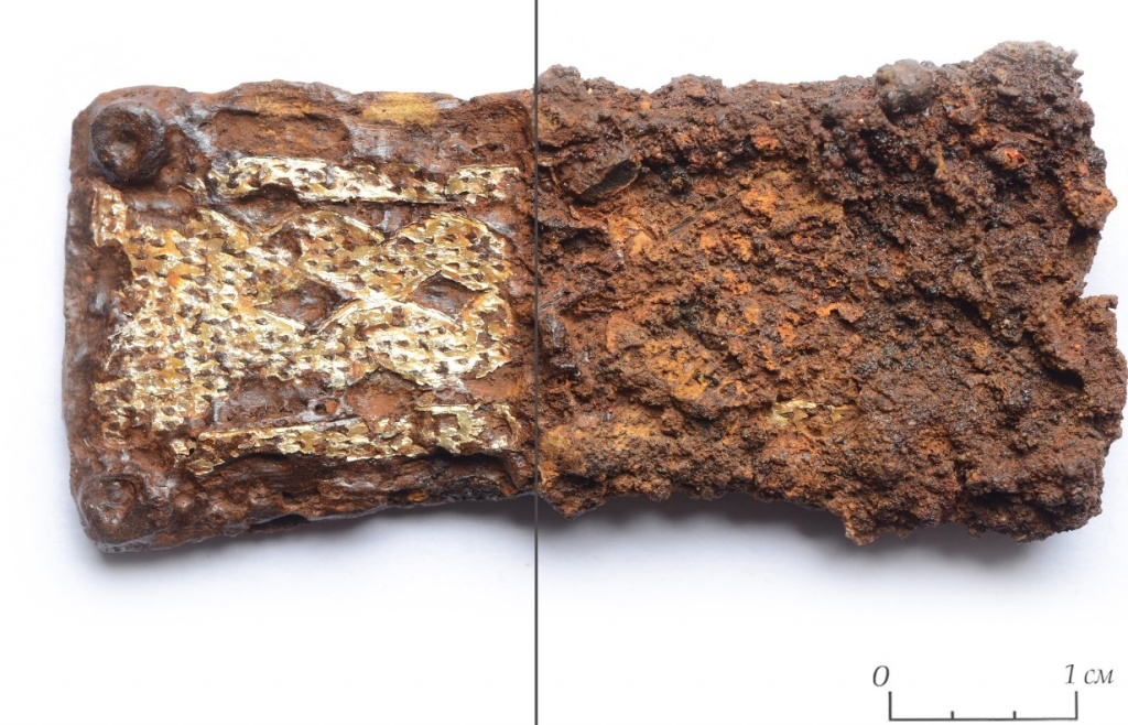
Fig. 13. Harness belt onlay. On the right – mineralized iron layers conceal the ornament completely. On the left – inlay revealed by restoration
Analysis of the distribution maps of elements makes it possible to determine the boundaries of the ornament invisible to the naked eye. The reason for it is corrosion layers being somewhat transparent to X-ray. Thus on the map of iron distribution (Fig. 11) the presence of the ornament is not noticeable, because the ornament is hidden by layers of mineralized iron, which is obvious during visual inspection. However, on the map of distribution of silver (Fig. 4) ornament borders made with the precious metal on iron can be easily identified. This is inlaid silver foil on an iron base. Resolution of the microscope allows to partially see the traces of a peculiar method of fixing the silver foil to the iron base. Those are small points distributed over the surface of silver (Fig. 4), traces of holes punched by the chisel through the silver foil. After each strike, the foil was pressed into the iron and thus fixed on the buckle. After revealing restoration these holes are clearly visible in the image.
Mapping of other elements also provides meaningful information. Firstly, on the surface of the foil there is gold (Fig. 3). Its small amount is unevenly distributed over the surface. For a better understanding, local distribution of gilding layer on the ornament is shows very clearly on the combined gold and silver map (Fig. 11). Silver foil was apparently gilded to make an ornament of golden color that we see after the reveal. For restoration not only the presence of the ornament has to be determined, but traces of gilding before the start of the reveal are also of great importance. After all, the thinnest gilding, more so unevenly preserved, can be damaged during reveal, unless noticed in advance. Secondly, the presence of copper (Fig. 5) can be connected to using gold as raw material for gilding. Thirdly, the distribution of calcium and silicon (Fig. 6 and 7), can be attributed to contamination with soil, because the object is of archaeological origin. The rest of the elements can also be attributed to random impurities.
Microscope-microprobe XROS MF30 allows to conduct elemental analysis and elemental mapping of various archaeological sites without their destruction. The information obtained with it is extremely useful in the restoration of such objects.
The method of X-ray fluorescence mapping makes it possible to obtain maps of the element distribution, which can explain technical and technological features of archaeological objects made of metal, the composition of alloys used for the base and the ornament. It is very important that during the experiment it was possible to show the applicability of the method to detect the ornament invisible on the surface even before the restorative intervention. Since the method is nondestructive, it was done without any harm and without changes in the material structure of the object.
Obtaining such information by other methods is almost impossible. Radiographic studies are difficult due to the great thickness of the objects. Electron microscopy makes it impossible to obtain an elemental mapping of an object covered with a thick layer of corrosion, soil and mineralized organic matter due to the low penetrating power of electrons.
*[1] Prokuratov D. S., Parfenov V.A., Barrows N. Gorlov V.K., Grigorieva I.A., Urupov S.O. Application of Laser Technology in the Restoration of Archaeological Metals // Materials of the SpbPU Science Week Scientific Forum with International Participation Conference, Saint-Petersburg, 30 November-5 December 2015, 2015, pp. 112-114